
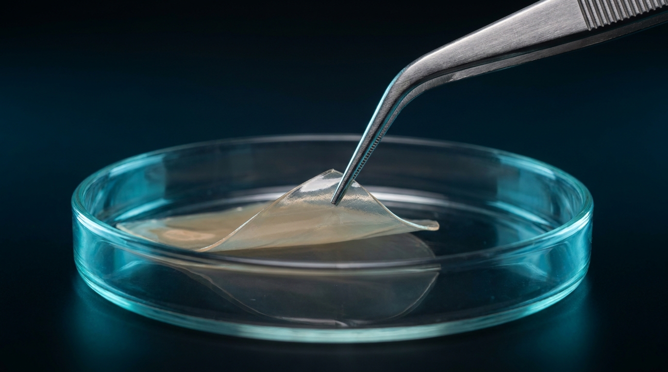
Where chitosan comes from.
After cellulose, chitin is one of the most abundant biopolymers (Younes & Rinaudo 2015). It forms the framework of crustacean shells, fungal cell walls and insect cuticles. Chitosan can be obtained from all three sources.
From raw material to Z-Chitosan: Origin & botanical actives
| Source | Raw material | Notes |
|---|---|---|
| Crustaceans | shrimp and crab shells, a by-product of the food industry | main source; over 80,000 t of chitin a year come from marine by-products (Ghormade et al. 2017) |
| Fungi | cell walls, e.g. of Aspergillus niger, Mucor rouxii or Rhizopus oryzae | more uniform chain length and high degree of deacetylation reported (Ghormade et al. 2017) |
| Insects | e.g. pupal exuviae of the black soldier fly from insect farming | up to 25.5 % chitin in the pupal exuviae (Triunfo et al. 2022) |
From chitin to chitosan.
01
Demineralisation
dilute hydrochloric acid (typically 0.25–2 mol/l) dissolves the calcium carbonate of the shell, releasing CO₂.
02
Deproteinisation
sodium hydroxide solution (typically 0.3–2.5 mol/l, 65–100 °C) removes the proteins, leaving chitin.
03
Deacetylation
concentrated sodium hydroxide at elevated temperature removes acetyl groups, turning chitin into chitosan.
Temperature, alkali concentration and time determine the degree of deacetylation and the molar mass (Younes & Rinaudo 2015).
Key properties.
| Parameter | Typical value | Significance |
|---|---|---|
| Structure | β-(1→4)-linked D-glucosamine and N-acetyl-D-glucosamine | linear polysaccharide |
| Degree of deacetylation (DDA) | typically 60–95 % | share of free amino groups, sets the charge density |
| Molar mass | in studies e.g. 60–1,370 kDa (Wang et al. 2006) | affects viscosity, film formation and activity |
| pKa of the amino groups | about 6.2–6.5, depending on DDA and molar mass (Wang et al. 2006) | below it, the charged form –NH₃⁺ dominates |
| Solubility | soluble in dilute acids, barely at neutral pH (Rinaudo 2006) | usually processed from acidic solution |
Charge across the pH range.
The Henderson–Hasselbalch equation estimates how many amino groups are charged. With a pKa of 6.4, about 96 % are charged at pH 5 and only about 20 % at pH 7. This is a simplified model; in polyelectrolytes the apparent pKa shifts with the degree of charge.
88,8 % protonated
Protonated share = 1 / (1 + 10^(pH − pKa))
What am I looking at?
| pH | protonated (–NH₃⁺) | uncharged (–NH₂) |
|---|---|---|
| 3.0 | 99.96 % | 0.04 % |
| 4.0 | 99.6 % | 0.4 % |
| 5.0 | 96.2 % | 3.8 % |
| 6.0 | 71.5 % | 28.5 % |
| 6.4 | 50.0 % | 50.0 % |
| 7.0 | 20.1 % | 79.9 % |
| 7.4 | 9.1 % | 90.9 % |
| 8.0 | 2.5 % | 97.5 % |
| 9.0 | 0.3 % | 99.7 % |
How chitosan acts: what the literature discusses.
The mechanism is not fully understood. Reviews by Rabea et al. (2003) and Kong et al. (2010) describe several routes; their contribution depends on organism, molar mass and pH.
Charge: positively charged chains bind to negatively charged cell surfaces, to lipopolysaccharide in Gram-negative and teichoic acids in Gram-positive bacteria. The membrane becomes leaky and cell contents escape.
Chelation: chitosan binds metal ions needed by the cell envelope and enzymes.
Barrier: high-molar-mass chitosan can coat the cell with a film that hinders exchange.
Cell interior: for short chains, binding to DNA that disrupts mRNA synthesis is discussed. This route is disputed.
The model in 3D: Cell Envelope 360°
Literature
Known limits of plain chitosan.
Activity depends strongly on pH (Kong et al. 2010). At pH 7 the model gives only about one fifth of the amino groups a charge, and chitosan precipitates. Many settings, from plastic surfaces to blood, lie in this range.
Degree of deacetylation and molar mass depend on raw material and process (Younes & Rinaudo 2015), and both affect activity (Younes et al. 2014). Batches therefore need specifying and testing.
The known crustacean allergens are proteins, mainly tropomyosin (Reese et al. 1999); chitosan itself is a polysaccharide. In a small study, ten people with shellfish allergy tolerated a chitosan wound dressing without reaction (Waibel et al. 2011). The data are limited, so the source should be stated openly on every data sheet.
Susceptibility differs between species and growth phases (Kong et al. 2010). Results against one organism cannot be transferred to others.
Fungal or crustacean chitosan.
Both are the same type of polymer and differ in raw material, uniformity and the allergen question. The patent specifications for Z-Chitosan do not name a raw material source; information on the raw material source of Z-Chitosan is available on request.
| Feature | Crustacean chitosan | Fungal chitosan |
|---|---|---|
| Raw material | shrimp and crab shells | fungal biomass, e.g. from fermentation |
| Market position | established main source | complementary source (Ghormade et al. 2017) |
| Uniformity | varies with raw material and process | more uniform chain length reported |
| Allergen question | declare crustacean origin | no crustacean proteins |
What Z-Chitosan does differently.
Z-Chitosan is an inclusion complex of chitosan and at least one plant extract, formed in the presence of a catalyst (US 11,708,424). It is produced by multi-stage emulsion cross-linking and dried to a powder; as a masterbatch it goes into plastics, polymers and fabrics (US 12,071,490).
The combination of chitosan with plant extracts is described in US patent 11,708,424 B1, “Inclusion of chitosan and herbal extracts as an antimicrobial agent”, granted on 25 July 2023.
Testing on the specific material shows the resulting properties in use.
For your product
What this means for your product.
First establish the pH range your product works in. For neutral environments, the textbook activity of plain chitosan is not enough; you need measured data on the finished material. Also ask for a batch specification with source, degree of deacetylation and molar mass.