
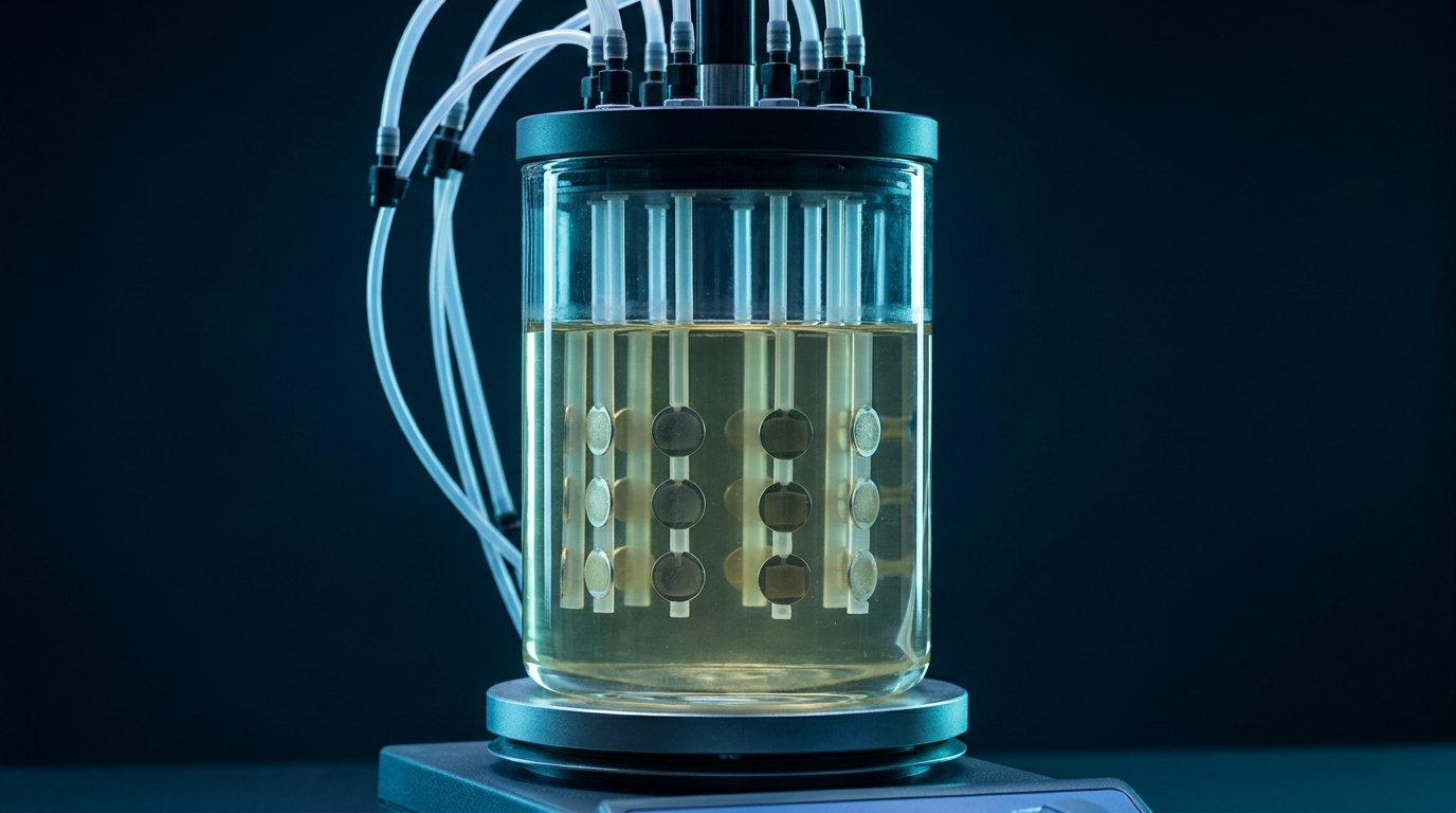
Why the start matters.
A biofilm is a community of microorganisms living in a self-produced matrix of extracellular polymeric substances (EPS) [1]. The matrix holds the cells together and hinders the penetration of active substances. Cells in a biofilm are markedly more tolerant of antimicrobial agents than free-floating cells [2].
On implants and catheters, a race for the surface begins: host cells and bacteria compete for the same space [3, 4]. Proteins from blood, urine or tissue fluid often adsorb first. This conditioning film changes the surface before the first cells arrive.
Effective strategies therefore act as early as possible. The longer a product stays in use, the more important it is that the effect lasts for its entire service life. The Biofilm Time Machine on the Medical devices page shows how biofilm forms in five phases [5, 6].
Literature
Four strategies against biofilm.
Contact killing
- Where it acts
- surface
- Kills
- yes
- Strength
- no release into the surroundings
- Limit
- direct contact only, covered by deposits
- Typical test
- ISO 22196, ASTM E2149
Contact killing. Firmly bound, mostly cationic groups disrupt the cell envelope of microorganisms that touch the surface [9]. No active substance is released. The effect reaches only as far as direct contact; dead cells and deposits can cover the active layer [10]. Fine nanostructures can also damage cells mechanically on contact [8].
Release killing
- Where it acts
- surface and surroundings
- Kills
- yes
- Strength
- also acts at a distance
- Limit
- finite reservoir, substance in the surroundings
- Typical test
- zone of inhibition test, ISO 22196
Release killing. The material releases an active substance, such as silver ions, an antiseptic or an antibiotic, which also acts in the immediate surroundings. The reservoir is finite and the amount released falls over time [11]. The substance reaches fluids, tissue or wastewater, which has to be considered in assessment and approval.
Preventing attachment
- Where it acts
- surface
- Kills
- no
- Strength
- nothing consumed, nothing released
- Limit
- defects, protein film
- Typical test
- adhesion assay, biofilm reactor (ASTM E2562)
Preventing attachment (anti-adhesive). The surface gives cells nothing to hold on to, for example through highly hydrated polymer brushes such as polyethylene glycol, zwitterionic layers or microstructures. Nothing is killed. Small defects or a protein film can cancel the effect over time [7, 8].
Antifouling, detachment
- Where it acts
- interface
- Kills
- no
- Strength
- also removes existing deposits
- Limit
- needs flow or an active agent on site
- Typical test
- flow or reactor model
Antifouling and detachment. Low-surface-energy surfaces, for example silicone-based ones, hold deposits so weakly that flow removes them (fouling release) [7]. Enzymatic approaches break down the matrix of an existing biofilm and release cells from it [12]. Both need shear forces or an active agent on site and do not kill by themselves.
Combinations. Many developments combine two strategies, for example an anti-adhesive layer with a contact-active or releasing component [13].
What am I looking at?
- Preventing attachment (anti-adhesive). The surface gives cells nothing to hold on to, for example through highly hydrated polymer brushes such as polyethylene glycol, zwitterionic layers or microstructures. Nothing is killed. Small defects or a protein film can cancel the effect over time [7, 8].
- Contact killing. Firmly bound, mostly cationic groups disrupt the cell envelope of microorganisms that touch the surface [9]. No active substance is released. The effect reaches only as far as direct contact; dead cells and deposits can cover the active layer [10]. Fine nanostructures can also damage cells mechanically on contact [8].
- Release killing. The material releases an active substance, such as silver ions, an antiseptic or an antibiotic, which also acts in the immediate surroundings. The reservoir is finite and the amount released falls over time [11]. The substance reaches fluids, tissue or wastewater, which has to be considered in assessment and approval.
- Antifouling and detachment. Low-surface-energy surfaces, for example silicone-based ones, hold deposits so weakly that flow removes them (fouling release) [7]. Enzymatic approaches break down the matrix of an existing biofilm and release cells from it [12]. Both need shear forces or an active agent on site and do not kill by themselves.
- Combinations. Many developments combine two strategies, for example an anti-adhesive layer with a contact-active or releasing component [13].
| Strategy | Where it acts | Kills | Strength | Limit | Typical test |
|---|---|---|---|---|---|
| Preventing attachment | surface | no | nothing consumed, nothing released | defects, protein film | adhesion assay, biofilm reactor (ASTM E2562) |
| Contact killing | surface | yes | no release into the surroundings | direct contact only, covered by deposits | ISO 22196, ASTM E2149 |
| Release killing | surface and surroundings | yes | also acts at a distance | finite reservoir, substance in the surroundings | zone of inhibition test, ISO 22196 |
| Antifouling, detachment | interface | no | also removes existing deposits | needs flow or an active agent on site | flow or reactor model |
Five steps to the right strategy.
01
Define the service life
hours (single-use product), days to weeks (catheter) or years (component, surface).
02
Describe the environment
wet or dry, flowing or stagnant, nutrients, temperature.
03
Define the contact
skin, mucosa, blood, food or technical media only.
04
Decide whether an active substance may leave the material. This determines whether contact or release systems are an option and shapes the regulatory route (Biocidal Products Regulation, MDR).
05
Draw up a test plan
one standard for surface activity and a biofilm model that resembles the application.
06
Set the success criterion before testing, for example the required log reduction compared with untreated material.
How anti-biofilm performance is tested.
ISO 22196 measures the antibacterial activity of a surface after 24 hours under a cover film at 35 °C and at least 90 % relative humidity [15]. The test shows killing or growth inhibition; it does not model a biofilm.
Biofilm has its own models. In the CDC biofilm reactor to ASTM E2562-22, a Pseudomonas aeruginosa biofilm (ATCC 700888) grows on coupons for 48 hours under high shear. The result is reported as log₁₀ CFU per unit area [14].
Since 2023, plastics and other non-porous surfaces have had a standard of their own. ISO 4768 grows a Staphylococcus epidermidis biofilm (ATCC 35984) on the sample for 48 hours at 35 °C, stains it with crystal violet and compares the absorbance with untreated material. Textiles and photocatalytic materials are excluded [16].
ISO 22196:2011 · ISO 4768:2023 · ASTM E2562-22
Four common mistakes in anti-biofilm claims.
- Using a result from a 24-hour surface test as a biofilm claim. Killing under a film is not the same as a grown biofilm.
- Testing only one organism although several occur in the application, such as Gram-positive and Gram-negative bacteria or yeasts.
- Testing only fresh material. Ageing, cleaning, sterilisation or contact with body fluids can change the surface.
- Testing without a conditioning film. In protein, serum or urine solution a surface behaves differently from pure buffer.
For your product
What this means for your product.
For catheters, tubing and other medical devices that remain in place, the chosen strategy determines the test plan and the regulatory route. Z-Chitosan combines a cationic biopolymer with plant extracts; whether it acts through contact, release or both in a given material is tested for each material.